Recent Publications (Original)
Quantitative 3D correlative light and electron microscopy of organelle association during autophagy (Takahashi et al., Cell Struct Funct.)
2022.12.22 Recent Publications (Original)
Satoru Takahashi, Chieko Saito, Ikuko Koyama-Honda*, Noboru Mizushima* (*co-corresponding authors)
Quantitative 3D correlative light and electron microscopy of organelle association during autophagy
Cell Structure and Function 47: 89–99 (2022) https://doi.org/10.1247/csf.22071
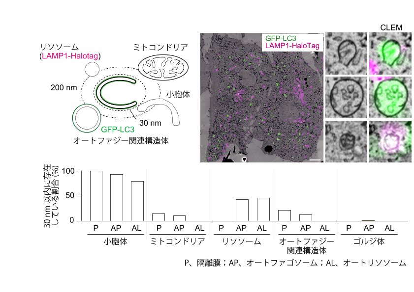
In macroautophagy, disk-shaped double-membrane structures called phagophores elongate to form cup-shaped structures, becoming autophagosomes upon closure. These autophagosomes then fuse with lysosomes to become autolysosomes and degrade engulfed material. Autophagosome formation is reported to involve other organelles, including the endoplasmic reticulum (ER) and mitochondria. Organelles are also taken up by autophagosomes as autophagy cargos. However, few studies have performed systematic spatiotemporal analysis of inter-organelle relationships during macroautophagy. Here, we investigated the organelles in contact with phagophores, autophagosomes, and autolysosomes by using three-dimensional correlative light and electron microscopy with array tomography in cells starved 30 min. As previously reported, all phagophores associate with the ER. The surface area of phagophores in contact with the ER decreases gradually as they mature into autophagosomes and autolysosomes. However, the ER still associates with 92% of autophagosomes and 79% of autolysosomes, suggesting that most autophagosomes remain on the ER after closure and even when they fuse with lysosomes. In addition, we found that phagophores form frequently near other autophagic structures, suggesting the presence of potential hot spots for autophagosome formation. We also analyzed the contents of phagophores and autophagosomes and found that the ER is the most frequently engulfed organelle (detected in 65% of total phagophores and autophagosomes). These quantitative three-dimensional ultrastructural data provide insights into autophagosome-organelle relationships during macroautophagy.
![]()
